Product
다올팬텀 제품을 소개합니다.
Radiography
| 제목 | Contrast-Detail-Phantom CDRAD |
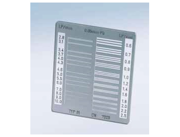
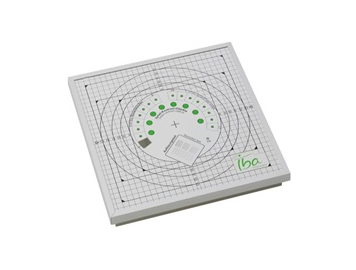
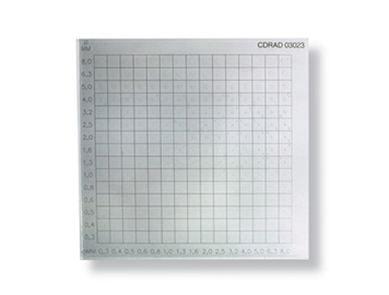  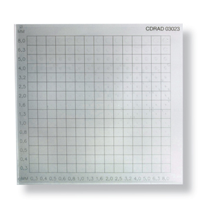 X-ray 장비의 대조도 팬텀 Contrast-Detail-Phantom CDRAD 입니다. 소프트웨어를 통해 엑스레이 장비의 대조도 분해능력을 평가할수 있습니다. 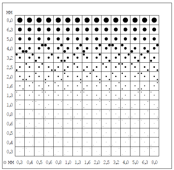 Introduction Most definitions of image quality in radiology are based on characterizing the physical properties of the image chain. However, medical diagnosis is not made by the image alone. The perception by the observer is crucial for the result. A test of the observers perception is possible with so called Contrast-Detail (CD) phantoms. With a CD-phantom it is possible to quantify both, detail and contrast, as observed by the radiologist. The CDRAD 2.0 phantom can be used within the entire range of diagnostic imaging systems, such as fluoroscopy and digital subtraction angiography. Applications Determination of the optimum exposure technique Optimization and evaluation of digital radiology systems Comparison of the image quality at various thicknesses of PMMA Comparison of the image quality versus dose relation Determination of the optimum background density Comparison of different radiology systems Features Aid for improving image quality Monitors the image information content Contrast-Detail curve/detectability Tests low contrast and spatial resolution Characteristics Automatic scoring diagram Averaging the score of multiple images Contrast Detail curve Image quality figure and % detected dots Statistically sound User definable settings Formats DICOM 3.0 Monochrome 1 and 2 Bitmap, jpeg and tiff files Runs on Windows XP®/ Vista®/ 7® platforms Free USB port needed 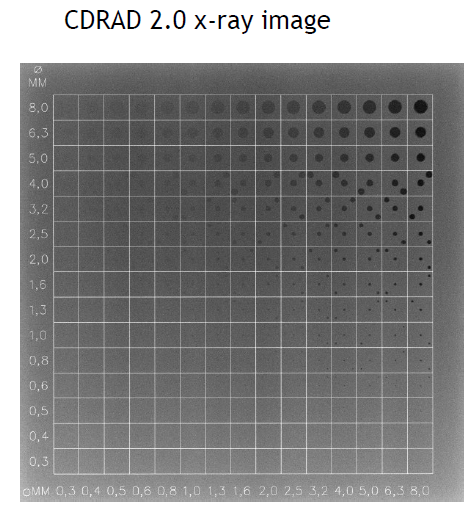 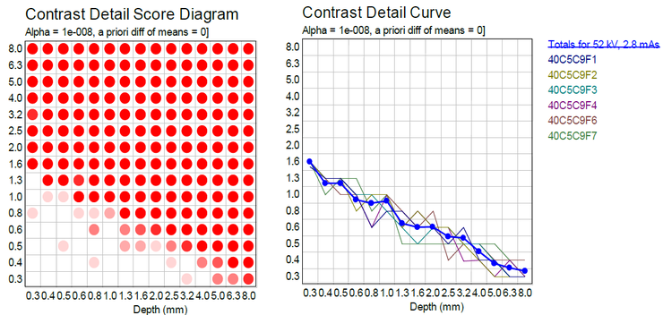 |
|
| 카테고리 | |
| 문의하기 |


|

|