특수의료장비 품질관리검사의 기준(제5조제2항 관련)
라. 혈관조영장치
표준팬텀
| Standard XR-21, National Electronical Manufacturers Association; NEMA
|
팬텀 촬영 조건
| 1. SID 100cm으로 설정
2. 80kVp, 20mA. 장치의 구비 조건상 기본촬영조건을 정확히 준수 할 수 없는 경우 가장 근접한 조건으로 검사하고 이를 명시함
3. 팬텀과 디텍터간의 거리는 50mm로 설정
4. C-arm의 RAO/LAO 및 CRA/CAU을 0도로 설정
5. 해상력 챠트는 모니터 주사선 및 그리드의 영향을 최소화하기 위하여 45도로 위치
6. 환자 테이블 위의 패드를 제거함
|
제출영상
| 1. 팬텀 촬영 스팟 영상 1부(영상에는 장비고유번호와 촬영날짜를 표시)
2. 고정식 이중면(dual)의 경우 양면에서 모두 검사하여 제출한다.
|
합격기준
| 이중면의 경우 양면에서 모두 적합이어야 적합이다.
|
항 목
| 합 격 기 준
|
Beam alignment
| Acceptable 기준 이상이어야 한다.
|
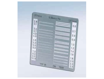
Spatial resolution
| I.I type
field size(cm)
| 34-40
| 0.8(lp/mm)
|
28-33
| 1.0(lp/mm)
|
22-27
| 1.2(lp/mm)
|
16-21
| 1.4(lp/mm)
|
10-15
| 1.5(lp/mm)
|
F.D type
field size(cm)
| Over 45
| 0.8(lp/mm)
|
40-45
| 1.0(lp/mm)
|
28-33
| 1.2(lp/mm)
|
22-27
| 1.4(lp/mm)
|
16-21
| 1.5(lp/mm)
|
Low contrast iodine detectability
| 최소농도 200mg/cc까지 보여야 한다.
|
Phantom
entrance dose
| 10R/min 이하이어야 한다. (화면 확대 비적용)
|
Wire resolution
| Static
| 최소 0.012“까지 보여야 한다.
|
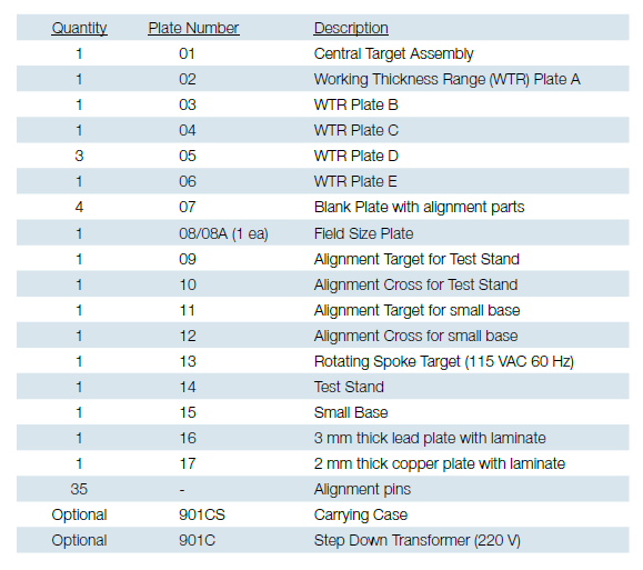
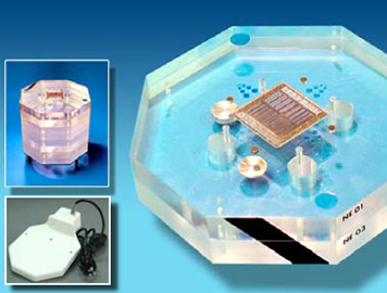
The CIRS Model 901NEMA-SCA&I phantom was designed to evaluate and standardize catheterization image quality. It is the result of collaborative efforts between the Society for Cardiac Angiography and Interventions and the National Electric Manufacturers Association.
The phantom specifically enables voluntary compliance with the recently published performance standard NEMA XR21.
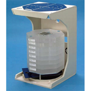
The Model 901 is manufactured from PMMA with X-ray absorption properties similar to soft tissue at diagnostic energies. It contains a variety of static and dynamic test targets for objective assessment of resolution, motion unsharpness and radiation exposure. The sectional design allows for configuration in a wide range of thicknesses from 5 to 30 cm simulating PA thicknesses from infants to large adult patients.
The phantom is ideal for routine assessment of the entire imaging system.