Product
다올팬텀 제품을 소개합니다.
Dental
| 제목 | DICOM기반 주문제작 CT팬텀 HEAD AND NECK PHANTOM |
 HEAD AND NECK PHANTOM FOR CT, X-RAY AND RADIATION THERAPY 환자의 DICOM파일을 실제와 거의 동일하게 제작하는 팬텀입니다. 고객이 원하는 DICOM파일을 전달해주시면 원하는 부위를 원하는 크기로 제작 할 수 있습니다. 다양한 DICOM 데이터를 이용하여 소아 영아 노인 등등 다양한 제작이 가능합니다. Lesion 이나 Low Contrast 등도 추가가 가능하며 여러 형태로 커스텀 주문제작도 가능합니다. 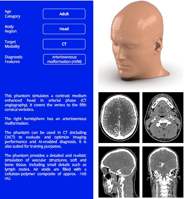 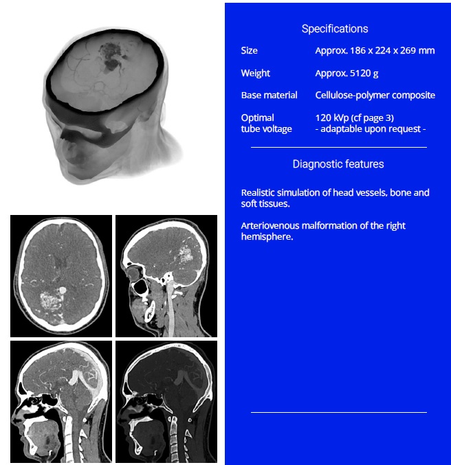 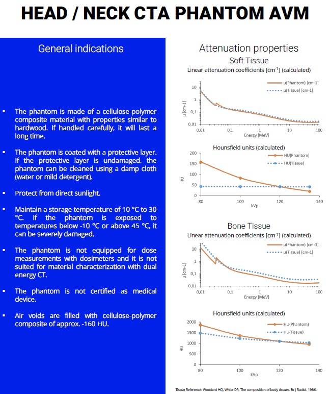 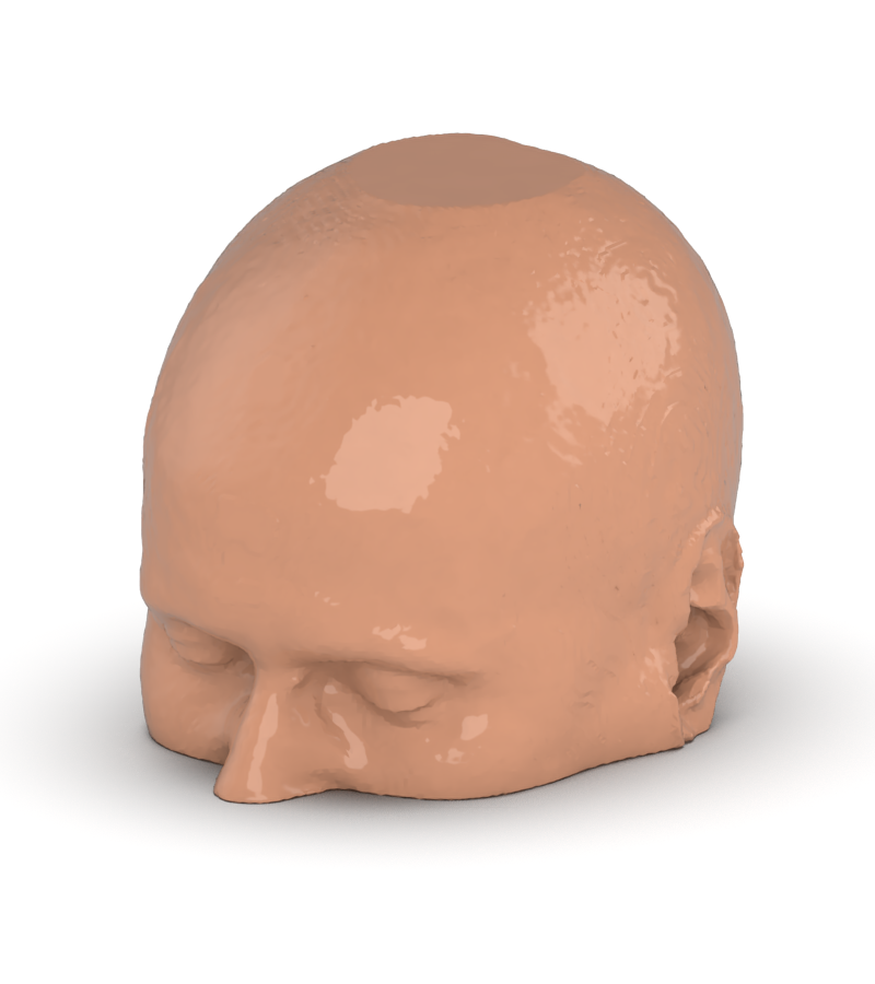 원하는 부위만 제작도 가능합니다. 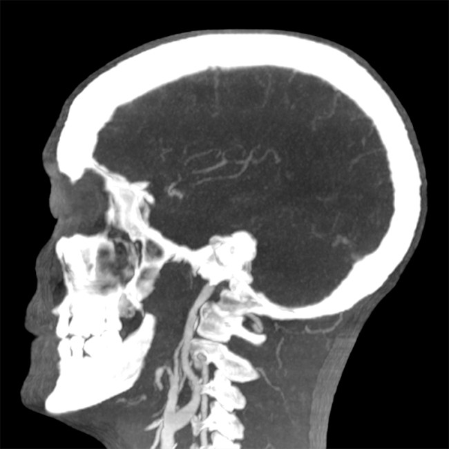 Product information "Head and Neck Phantom for CT, X-ray and Radiation Therapy" Head and neck phantom with realistic anatomy. This highly realistic head and neck phantom was designed to simulate clinical imaging and dose exposure in computed tomography including dual energy CT, X-ray imaging and radiation therapy. The model provides a realistic simulation of all tissues and realistic attenuation values. This phantom provides a detailed simulation of patient exposure and provides new opportunities for testing and optimizing image quality and dose, dose verification at low and high energy exposure and for training of medical and technical staff. The phantom is manufactured based on a real CT data set and includes anatomic details for all tissues. The model is a handmade unique piece, which can differ slightly in size and design. The phantom can be provided as one-piece anthropomorphic phantom or in a sectional design and it can include openings for dosimeters. Pathologic features (e.g., masses, vascular pathologies) can be included upon request into the phantom. 인체의 머리와 목 부위의 해부학적인 구조를 사실적으로 구현한 제품입니다. 일반적인 뼈를 보는 팬텀이 아닌 방사선 피폭에 따른 상세한 시뮬레이션이 가능하도록 구현된 팬텀입니다. 실제 CT장비에서의 데이터를 기반으로 만들어졌기 때문에 낮은 에너지 혹은 높은 에너지에서도 선량 검증과 트레이닝이 가능합니다. 수제로 작업하여 제작되기 때문에 디자인은 제품마다 조금씩 다를 수 있으며 선량계를 삽입할 수도 있는 개구부 또는 종양, 혈관 등의 병리학적 요소등을 넣을 수도 있습니다 |
|
| 카테고리 | 인체모형 |
| 문의하기 |


