Product
ДйПУЦвХв СІЧАРЛ МвАГЧеДЯДй.
CT/MRI
| СІИё | Triple Modality 3D Abdominal Phantom, model 057A |
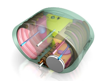
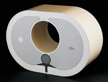
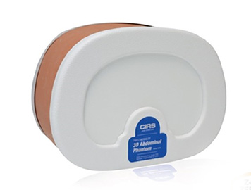  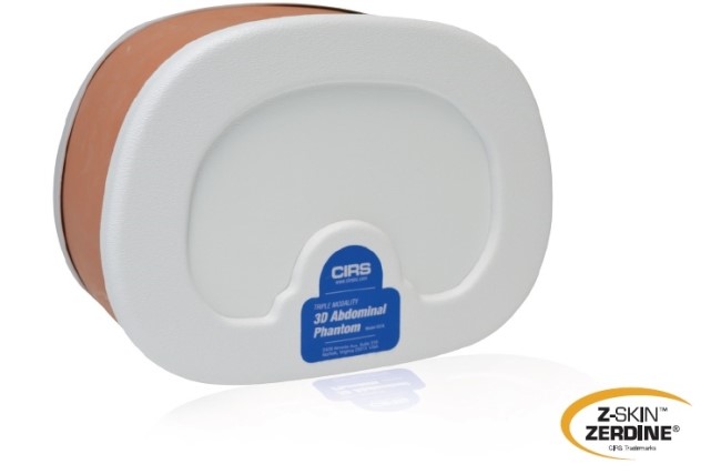 CT/ ULTRASOUND/ MRI IMAGE FUSION• LIVE SCANNING• BIOPSY TRAINING The CIRS Triple Modality 3D Abdominal Phantom is constructed of a self-healing formulation of ZerdineЂч(1) that allows multiple biopsy insertions with minimal needle tracking, and is ideal for demonstrating image-guided navigation technologies. Abdominal imaging is useful for diagnosing disease and monitoring treatments. The Model 057A is representative of a small adult abdomen and can be imaged under CT, MR and ultrasound. This feature makes the phantom a useful tool for applications such as image fusion studies; imaging protocol developments; scan technique training; and system testing, validation and demonstration. The Model 057A simulates the abdomen from approximately the thorax vertebrae (T9/T10) to the lumbar vertebrae (L2/L3) using simplified anthropomorphic geometry. The materials provide contrast between the structures under CT, MR and ultrasound. The solid polymer background gel will not leak when punctured.* Internal structures include the liver, the portal vein, two partial kidneys, a partial lung, the abdominal aorta, the vena cava, a simulated spine and six ribs. The liver has six lesions and the kidneys each have one lesion. A muscle layer and outside fat layer surround these structures and plastic end caps make the phantom durable enough for extended scanning. Blood vessels have CT contrast added to provide enhanced auto registration in image fusion applications The phantom includes a foam lined hard carry case. For users interested in image fusion studies, the phantom can be purchased as a kit to include a serial-number specific CT DICOM Data set for reference. CIRS can also offer value-added options and services such as phantom specific CMM, reference CT or MRI data sets, attachment of customer specific registration devices and inclusion of special point markers. 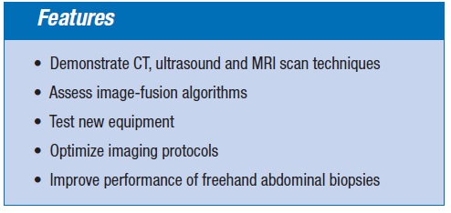  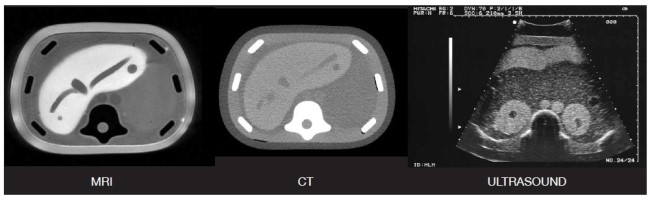 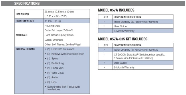 (1) US Patent # 5196343 (2) DICOM Images are provided with a free DICOM reader (Onis 2.6). If using alternate software to read the images, please notify CIRS of any special requirements for making the data compatible with your software. For instance, some programs include special checks of the DICOM header file or the DICOM directory when loading the image data set. 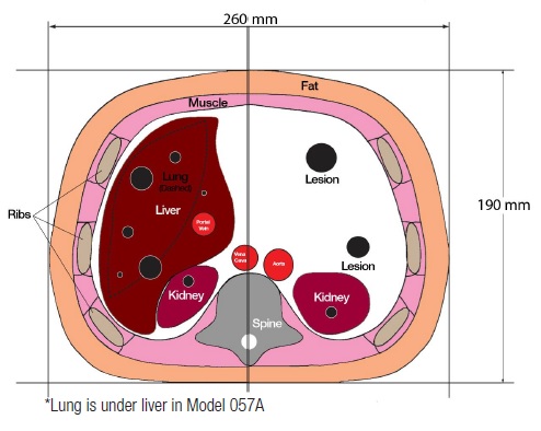 |
|
| ФЋХзАэИЎ | |
| ЙЎРЧЧЯБт |


|

|